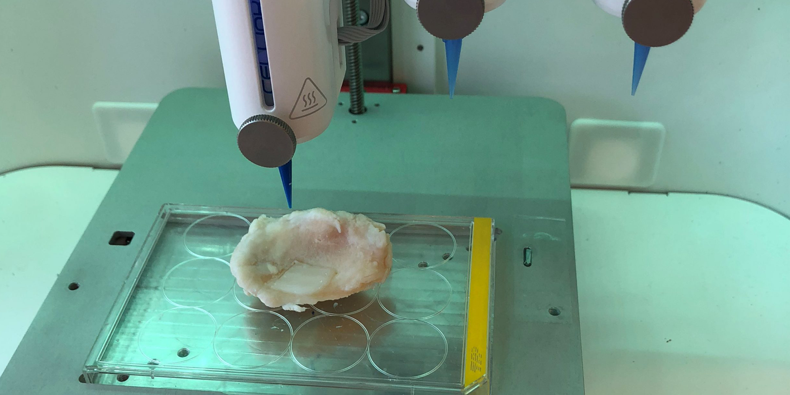

Recent article with the title “Collagen 2A Type B Induction after 3D Bioprinting Chondrocytes In Situ into Osteoarthritic Chondral Tibial Lesion” published by members of RESTORE (Cartilage, published online ahead of print 2020 Feb 18). It was shown that cartilage could be bioprinted in three-dimensions (3D) to match an injury of the tibia. Tibia is the front bone below the knee and articular cartilage is the smooth ending of the bones in the joint. Osteoarthritis (OA) disease develops slowly and cartilage is degraded in several stages before it ends up in total joint destruction and the most common treatment is joint replacement. If the OA-degeneration process of cartilage is interfered with so it slows down, it might postpone joint replacement. Local cartilage repair using autologous chondrocyte implantation (ACI) has previously been shown to halt such a degenerative process. For more than 30 years, local traumatic cartilage lesions have been treated using ACI. In this study, an OA defect site was scanned using magnetic resonance imaging (MRI) and computer tomography (CT), microcomputer tomography (MicroCT) and a 3D scanner to obtain a 3D model of the defect for 3D bioprinting using primary chondrocytes obtained after ACI surgery. Chondrocytes are the only cells found in articular cartilage and has previously been shown to contain high levels of collagen IIA splice variant type B and collagen II (both type A and B) is undetectable and after culturing in monolayer.
The results showed that 3D scanner was the only device (except for microCT) that detected the OA defect area. Cartilage tissue that matched the OA lesion was 3D bioprinted. In support that mature articular cartilage can be formed after 3D bioprinting the authors showed that Collagen II A isoform type B levels (analysed by RT-PCR) was surprisingly higher in chondrocytes retrieved from the 3D bioprinted tissue than compared to control micromass. The ink used supported also the primary chondrocytes to produce ACAN, which is also a gene characteristic of articular cartilage. It was concluded by using 3D bioprinting technology a tissue engineered construct with cartilage repair capacity that match a defect could be produced.
More information:
https://journals.sagepub.com/doi/10.1177/1947603520903788
3D bioprinting in situ into an Osteoarthritic cartilage lesion
May 6, 2020